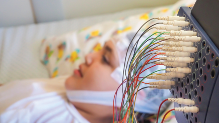
MEG
MEG (magnetoencephalography) provides a noninvasive tool to study epilepsy and brain function. When it is combined with structural imaging, it is known as magnetic source imaging (MSI).
- MEG measures small electrical currents arising inside the neurons of the brain. These currents produce small magnetic fields. MEG generates a remarkably accurate representation of the magnetic fields produced by the neurons.
- To some ways, MEG is similar to EEG (electroencephalography).
- An important difference is that the skull and the tissue surrounding the brain affect the magnetic fields measured by MEG much less than they affect the electrical impulses measured by EEG. This makes the MEG more accurate than an EEG in some ways. The test can give more usable and reliable information about the location of brain function.
- When MEG is added to magnetic resonance imaging (MRI) (which shows brain structure) the combination of the images is extremely helpful. Areas of the brain that could generate seizures as well as normal electrical activity in the brain can be located more easily.
Why Is An MEG Performed?
In the evaluation of epilepsy, MEG is used to localize the source of epileptiform brain activity, which most likely is the source of seizures. It is usually performed with EEG at the same time.
MEG may be helpful in the following situations:
- It can improve the detection of potential sources of seizures by revealing the exact location of the problem. This can help doctors find the cause of the seizures.
- It can help when MRI scans show a lesion or spot, but the EEG findings give different information. An MEG may be able to confirm that the epileptiform discharges (the brain waves typical of epilepsy) are indeed arising from the lesion. Then a decision can be made regarding surgery.
- In patients who have brain tumors or other lesions, the MEG may be able to map the exact location of the normally functioning areas near the lesion. Surgery to remove the tumor can be planned to lessen postoperative weakness or loss of brain function.
- In patients who have had past brain surgery, the electrical field measured by EEG may be distorted by the changes in the scalp and brain anatomy. If further surgery is needed, MEG may be able to provide necessary information without invasive EEG studies.
Preparing For The MEG Procedure
- No special preparations are needed for an MEG, unless sedation is planned.
- If sedation or other medicines are given before the test, you (or your child) may be asked not to eat after midnight.
- Regular medicines should be taken with a little bit of water.
- Wear loose, comfortable clothing.
- Do not wear jewelry, hair spray, make-up, hearing aids, or removable dental work.
- If you have a vagus nerve stimulator (VNS) or pacemaker, you may not be able to have MEG. Ask your doctor.
What will happen in the MEG lab?
- You will be asked a series of medical questions to make sure that your body does not contain any metallic objects that may interfere with the MEG.
- A videotape eraser will be moved over your head to erase magnetic activity from fillings in your teeth. You will also be asked about any previous surgeries.
- You will need to remove all clothing that has metal (such as zippers, snaps, or sparking paint) and change into a hospital gown or pants.
- EEG electrodes will be glued all around your head and one will be placed over your heart. Three small coils will be taped to your forehead and you will wear two other coils attached to earplugs.
- You will be asked to lie down on an MEG bed, where a small metal coil will touch all the different dots around your head to record its shape, and this information will go into the computer.
- After your head shape has been recorded in the computer, you will get ready for the MEG study itself. You may be given pillows to put under your knees and elbows, and blankets to keep you warm and comfortable. The sensors will be put over your head but will not cover your face. The coils and EEG electrodes will be plugged into the sensors.
- When you are comfortable, your family and the technologist will leave the room and the door will be closed. Closing the door can be a little scary, but you will have a small microphone so you can talk to the technologist, who can come into the room at any time.
- The MEG test will take between 1 hour and 2 1/2 hours. During this time, you will need to remain as still as possible, not moving your head. This is very important. If you need a break, tell the technologist.
- Sometimes stimulation tests are done. If you have this kind of test, little plastic sensors may be placed on your fingers, or you will be shown a video with different colors. By doing this test, the doctors will know which part of your brain controls your hands, feet, and vision.
- When all the testing is done, you will be removed from the MEG room. You can change back into your regular clothes and go home.
- Technicians and doctors will later review the information and report the results to your referring doctor.
Resources
Epilepsy Centers
Epilepsy centers provide you with a team of specialists to help you diagnose your epilepsy and explore treatment options.
Epilepsy Medication
Find in-depth information on anti-seizure medications so you know what to ask your doctor.
Epilepsy and Seizures 24/7 Helpline
Call our Epilepsy and Seizures 24/7 Helpline and talk with an epilepsy information specialist or submit a question online.
Tools & Resources
Get information, tips, and more to help you manage your epilepsy.


